What is DCIS?
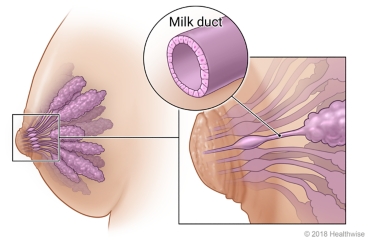
Ductal carcinoma in situ (DCIS) is the growth of abnormal cells in the milk ducts of the breast. It's an
early form of noninvasive breast cancer. Noninvasive means that the cells haven't spread. Some cases of DCIS
will become invasive breast cancer, but it's impossible to know which ones.
What causes it?
The exact cause of DCIS isn't known. Age and family health history may play a part.
What are the symptoms?
Most of the time, DCIS doesn't cause symptoms. But in some cases, symptoms can include a lump in the breast
or fluid or blood coming from the nipple.
How is it diagnosed?
DCIS is usually seen as small bits of calcium (microcalcifications) on a mammogram. To diagnose DCIS, your
doctor will remove a sample of breast tissue and look at it under a microscope. This is called a breast
biopsy.
How is it treated?
Treatment for DCIS is based on the grade and location of the cancer and other things, such as your overall
health and what matters to you. The main treatment is:
- Surgery.
- The choices are:
-
Breast-conserving surgery. This removes just the cancer and a border of healthy tissue around it.
-
Mastectomy. This removes the whole breast. Nearby tissue may also be removed and checked for cancer
cells.
-
Other treatments may include:
- Radiation therapy.
- This uses high-dose X-rays to destroy cancer cells and shrink tumors. Radiation may be given after
surgery. - Endocrine therapy.
- These medicines block hormones that cause certain cancers to grow. This helps slow or stop cancer growth.
Clinical trials are being done to find out if active surveillance may be an option for some people with DCIS.
Your doctor will talk with you about your options and then make a treatment plan.
Follow-up care is a key part of your treatment and safety. Be sure to make and go to all
appointments, and call your doctor if you are having problems. It's also a good idea to know your test results
and keep a list of the medicines you take.
Current as of: October 25, 2023
Content Version: 14.0
Care instructions adapted under license by your
healthcare professional. If you have questions about a medical condition or this instruction, always ask
your healthcare professional. Healthwise, Incorporated disclaims any warranty or liability for your use of
this information.
Featured in
Personalized care starts with the right provider
Search by specialty, location, and more.
FIND A DOCTORRelated Articles
Cystography
Cystography is an imaging test that can help diagnose problems in your bladder. It uses X-rays. They may be X-ray pictures or fluoroscopy, a kind of X-ray "movie."
Endometrial Cancer: Diagnosis
Cancer of the endometrium is a disease in which cancer cells are found in the lining of the uterus. It is highly curable when found early.
Normal Breast Development and Changes
Breast development happens in certain stages during a woman's life: first before birth, and again at puberty and later during the childbearing years. Changes also happen to the breasts during menstruation and when a woman reaches menopause.
Infectious Diseases
Detailed information on infectious diseases in children